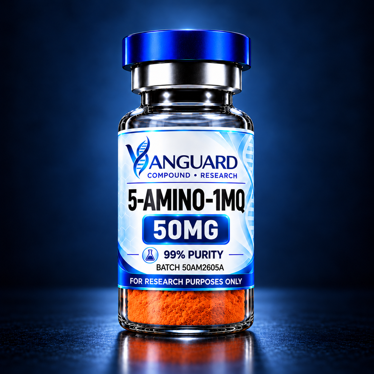
KPV is a short tripeptide composed of lysine, proline, and valineand corresponds to the C-terminal three-amino-acid sequence of alpha-melanocyte-stimulating hormone (α-MSH). It is primarily investigated for its potential role in inflammatory signaling, epithelial protection, immune regulation, and tissue-response pathways. Unlike full-length α-MSH, KPV retains substantial anti-inflammatory activity in experimental models while lacking its pigment-producing effect. Research has examined KPV in intestinal epithelial cells, immune cells, skin models, and airway epithelial systems, with particular interest in NF-κB and MAPK signaling. Experimental studies have also investigated its effects on pro-inflammatory cytokine production, oxidative stress, and cellular responses to inflammatory injury. Because of its very small three-amino-acid structure, KPV is useful for studying how minimal peptide sequences can retain specific biological signaling properties of larger parent peptides. Most evidence remains preclinical or mechanistic, and KPV has not been established as an approved therapeutic treatment. This material is intended for laboratory research only, is not authorized by Health Canada for human therapeutic use, and is not intended for human or veterinary administration.
Choose a kit option to save more, or skip this and buy individually above.


*LC-MS identity confirmation may be performed when analytical review indicates conflicting, atypical, or inconclusive results.
KPV, short for Lysine–Proline–Valine, is a synthetic tripeptide corresponding to residues 11–13 of alpha-melanocyte-stimulating hormone (α-MSH).
Its sequence is:
Lys – Pro – Val
or simply:
KPV
Despite consisting of only three amino acids, KPV has attracted considerable research interest because it retains much of the anti-inflammatory activity associated with α-MSH while lacking the pigmentary activity characteristic of the full peptide. PubMed
KPV is therefore commonly studied in experimental models involving:
Lysine forms the N-terminal amino acid of the KPV sequence.
Lysine is a basic amino acid containing an additional positively charged amino group on its side chain under physiological conditions.
Within peptides, lysine can influence:
In KPV, lysine contributes to the peptide's overall physicochemical character and may influence how the small peptide interacts with transport proteins and intracellular molecular targets.
Proline is structurally unusual among amino acids because its side chain loops back and bonds to its own amino nitrogen.
This cyclic structure restricts molecular flexibility and can influence peptide conformation.
Proline is frequently important in small bioactive peptides because it can:
Within KPV, proline helps create the compact structural geometry characteristic of the tripeptide.
Valine is a branched-chain amino acid and forms the C-terminal residue of KPV.
Its hydrophobic side chain contributes to molecular interactions involving nonpolar regions of proteins and cellular structures.
Within the KPV sequence, valine completes the minimal C-terminal fragment derived from α-MSH.
Together, lysine, proline, and valine form a remarkably small peptide capable of retaining biological effects associated with the much larger parent hormone.
Alpha-melanocyte-stimulating hormone is a peptide derived from the larger precursor protein proopiomelanocortin, or POMC.
α-MSH is associated with several biological processes, including:
Researchers discovered that the C-terminal KPV sequence retained substantial anti-inflammatory activity even though it lacked the full sequence required for α-MSH's pigmentary effects.
This finding made KPV especially interesting as a research model for separating the anti-inflammatory biology of α-MSH from its melanogenic actions. PubMed
The primary area of KPV research is inflammatory regulation.
Experimental studies have shown that KPV can influence intracellular signaling pathways involved in inflammatory responses.
One important pathway is:
NF-κB
NF-κB is a transcription factor that regulates expression of numerous inflammatory genes.
When activated, NF-κB can increase production of inflammatory mediators and cytokines.
KPV has been shown in experimental systems to suppress NF-κB activation and reduce production of certain inflammatory signaling molecules. PubMed
Another signaling system investigated with KPV is the MAP kinase pathway.
MAPK pathways help regulate cellular responses to:
In intestinal epithelial and immune-cell studies, KPV has been associated with suppression of inflammatory MAPK signaling along with reductions in cytokine production. PubMed
This makes KPV useful for studying how short peptides can influence multiple inflammatory signaling pathways simultaneously.
One of the strongest areas of preclinical KPV research involves the intestinal epithelium.
The intestinal lining is constantly exposed to:
Maintaining controlled inflammatory signaling is therefore essential for intestinal homeostasis.
Laboratory research has investigated KPV in intestinal epithelial cells and experimental mouse models of colitis.
Studies have reported reductions in inflammatory signaling and pro-inflammatory cytokine expression in these models. PubMed
A particularly interesting aspect of KPV biology involves the peptide transporter PepT1.
PepT1 normally transports small dipeptides and tripeptides across cellular membranes.
Researchers have demonstrated that KPV can be transported into intestinal epithelial and immune cells through PepT1.
This provides a useful mechanistic explanation for how such a small peptide may enter cells and influence intracellular signaling pathways.
Experimental studies found that PepT1-mediated KPV uptake was associated with inhibition of NF-κB and MAPK signaling. PubMed
Inflammatory responses rely heavily on signaling molecules known as cytokines.
KPV research has examined changes in several inflammatory mediators following cellular stimulation.
Experimental observations include reductions in signaling associated with:
These effects appear to occur upstream at transcriptional and signaling levels rather than simply neutralizing one individual cytokine.
KPV has also been investigated in skin biology, particularly in keratinocytes.
Keratinocytes form the majority of the outer epidermis and play an active role in immune and inflammatory signaling.
Experimental studies have examined KPV in models involving:
A 2025 keratinocyte study reported that KPV reduced oxidative-stress and inflammatory signaling in cells exposed to particulate matter, including effects involving reactive oxygen species and NF-κB-associated pathways. PubMed
These findings remain experimental and should not be interpreted as established clinical skin-treatment effects.
Oxidative stress occurs when production of reactive oxygen species exceeds normal cellular antioxidant defenses.
Reactive oxygen species can activate inflammatory pathways and contribute to cellular injury.
KPV has therefore been studied in models examining the relationship between:
oxidative stress → inflammatory signaling → cellular injury
Experimental keratinocyte research has shown reductions in reactive oxygen species and associated inflammatory signaling under specific laboratory conditions. PubMed
This provides another potential mechanistic research application for KPV beyond conventional cytokine biology.
KPV has also been studied in human bronchial epithelial cell models.
Research has examined its effect on inflammatory responses triggered by factors such as tumor necrosis factor and respiratory viral stimulation.
Observed experimental effects have included reductions in:
Researchers also investigated KPV's interaction with intracellular nuclear transport mechanisms involved in NF-κB signaling. PubMed
This work helps illustrate that KPV's research applications are not confined exclusively to intestinal tissue.
KPV has been investigated in immune-cell systems because α-MSH-derived peptides are associated with regulation of inflammatory responses.
Potential areas of investigation include:
The precise mechanism of KPV remains under investigation and does not appear to depend entirely on classical melanocortin-receptor signaling.
That distinction is important because KPV is substantially smaller than α-MSH and may interact with intracellular pathways differently. PubMed
Full-length α-MSH interacts with melanocortin receptors such as MC1R.
KPV, however, lacks much of the sequence normally associated with conventional melanocortin-receptor binding.
Research therefore suggests that at least some KPV activity may occur independently of classical α-MSH receptor signaling.
Studies in keratinocytes and inflammatory models have investigated alternative mechanisms involving:
This remains an active mechanistic research area. PubMed
Early experimental research has also examined KPV in host-defense models.
Laboratory studies involving α-MSH and KPV reported inhibitory effects against organisms including Staphylococcus aureus and Candida albicans under experimental conditions. PubMed
These observations are mechanistic and laboratory-based.
They should not be interpreted as evidence that KPV is an approved antimicrobial treatment.
PubChem identifies KPV as L-lysyl-L-prolyl-L-valine, molecular formula C₁₆H₃₀N₄O₄, molecular weight 342.43 g/mol, and CAS 67727-97-3. PubChem
KPV is a tripeptide, meaning it contains three amino-acid residues linked through peptide bonds.
Its sequence is:
H-Lys-Pro-Val-OH
The sequence corresponds to the C-terminal three residues of α-MSH.
Its small molecular size distinguishes it from many larger signaling peptides and makes it particularly useful for studying minimal bioactive peptide sequences. PubChem
Common research pathways include:
Research applications include:
Experimental applications include:
Research interests may include:
Experimental studies may evaluate:
A simplified experimental pathway can be represented as:
Inflammatory stimulus
↓
Cellular peptide uptake / signaling interaction
↓
NF-κB and MAPK pathway modulation
↓
Altered inflammatory gene expression
↓
Reduced inflammatory mediator production
Research indicates that several mechanisms may contribute, including peptide transport through PepT1 and intracellular effects on NF-κB-associated signaling. PubMed
Peptide length: 3 amino acids
Sequence: KPV
Full sequence: Lys-Pro-Val
Molecular formula: C₁₆H₃₀N₄O₄
Molecular weight: Approximately 342.43 g/mol
CAS number: 67727-97-3
Chemical class: Tripeptide
Nominal vial strength: 10 mg
Form: Lyophilized powder
Nominal content: 10 mg per vial
Compound type: Synthetic tripeptide
Primary research category: Inflammatory and epithelial signaling
Presentation: Research vial
Lyophilization is commonly used for research peptides to reduce water content and improve stability during appropriate storage before experimental preparation.
KPV is a small peptide and is commonly handled in aqueous laboratory systems.
Specific solvent selection, concentration, pH, and preparation procedures should be determined by the experimental protocol.
Relevant variables may include:
Consistency across these parameters is important when comparing experimental groups.
General laboratory handling considerations include:
Experimental storage conditions should be standardized within each research protocol.
KPV may be relevant to laboratory investigations involving:
Researchers may examine endpoints such as:
KPV 10 mg is intended for controlled laboratory investigation involving:
Most evidence supporting these research areas comes from cellular and animal models, and experimental findings should not automatically be extrapolated to therapeutic outcomes in humans. PubMed
KPV remains an investigational research peptide.
It is not authorized by Health Canada for human or veterinary use and is intended strictly for laboratory and research purposes.
It is not intended for clinical, diagnostic, therapeutic, or veterinary administration.
No documents available for this product yet.
Additional documentation available upon request.
Each batch undergoes analytical review using validated laboratory procedures. HPLC testing is utilized to assess purity and compare the sample profile against established reference standards. Where additional verification is warranted, LC-MS analysis may be performed to support compound identity confirmation.
*See LC-MS Confirmation Information for full details.
Every batch supplied by Vanguard Compound Research is assigned a unique batch number and maintained under documented quality-control procedures with supporting analytical documentation available upon request.
All Vanguard Compound Research orders are shipped directly from within Canada, ensuring efficient delivery, reliable service, and consistent quality-control from fulfillment to arrival.
Products are maintained under documented quality-control procedures to help ensure quality, consistency, and transparency.
Each product is assigned a unique lot number, allowing researchers to match their item directly to its corresponding Certificate of Analysis (COA).
Supporting analytical documentation may be available for qualifying products, including batch-specific quality records and testing information.
Documentation may include: